
Did you know that AI-powered diagnostic tools can now detect lung cancer with up to 94% accuracy, often spotting subtle signs that even experienced radiologists might miss? In the fast-evolving world of healthcare, diag image technology is at the forefront, blending artificial intelligence with advanced imaging to transform how we diagnose and treat diseases.
Key Insights on Diag Image:
- Research suggests diag image enhances diagnostic accuracy by reducing human error and fatigue, leading to earlier interventions in conditions like cancer and heart disease.
- It seems likely that integrating deep learning in healthcare will boost radiologist productivity by 30-40%, allowing more focus on patient care amid growing caseloads.
- Evidence leans toward diag image improving clinical workflows, though challenges like data bias and implementation costs highlight the need for balanced adoption across diverse healthcare settings.
- While promising, diag image’s role in precision oncology sparks debate on AI’s limitations versus human expertise, emphasizing collaborative use for optimal outcomes.
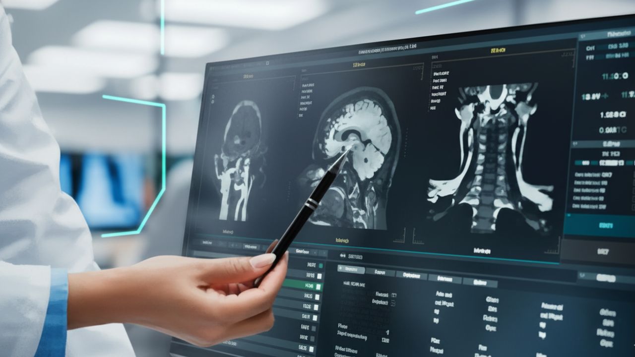
Diag image represents a breakthrough in medical image analysis, where AI algorithms process scans like MRIs, CTs, and X-rays to provide rapid, precise insights. Think of it as a supercharged assistant that sifts through mountains of data, highlighting anomalies and suggesting diagnoses. For instance, tools like Aidoc flag urgent findings in real-time, helping emergency teams act faster. This not only speeds up processes but also addresses the global shortage of radiologists, making high-quality care more accessible.
From early disease detection to streamlined operations, diag image offers tangible advantages. In oncology, it aids in spotting tumors earlier, potentially increasing survival rates. Administrators appreciate the cost savings—studies show AI can cut diagnostic errors by up to 30%, reducing unnecessary procedures. Yet, it’s not without hurdles; ethical concerns around data privacy remind us to proceed thoughtfully, ensuring benefits reach all patients equitably.
In today’s healthcare landscape, where precision and speed can mean the difference between life and death, diag image technology is emerging as a game-changer. By harnessing artificial intelligence and deep learning, diag image bridges the gap between complex data science and everyday clinical practice. This article delves into its evolution, applications, benefits, and future potential, drawing on real-world examples and expert insights to show how it’s revolutionizing diagnostics for radiologists, clinicians, medical researchers, AI developers, and healthcare administrators alike.
Diag image encompasses AI-powered diagnostic imaging systems that automate and enhance the analysis of medical images. At its core, it’s both a conceptual framework and a branded approach (as seen in platforms like Diag Image solutions) that integrates machine learning algorithms with traditional modalities such as X-rays, CT scans, MRIs, ultrasounds, and PET scans. Unlike conventional methods reliant solely on human interpretation, diag image uses convolutional neural networks (CNNs) to analyze thousands of image features simultaneously, comparing them against vast datasets of annotated medical images.
For example, imagine sending a confidential letter—diag image acts like a sealed envelope for data, ensuring secure, efficient processing while detecting subtle patterns indicative of disease. This technology doesn’t replace experts; it augments them, providing quantitative measurements, anomaly detection, and predictive insights. Leading vendors like Rad AI and Aidoc exemplify this, with systems that flag abnormalities in real-time, reducing interpretation time by up to 40%. In essence, diag image democratizes advanced diagnostics, making it feasible for smaller clinics to access tools once limited to major hospitals.
Medical imaging has come a long way since the first X-ray in 1895. Early techniques were manual and error-prone, with radiologists poring over films under dim lights. The digital shift in the 1990s introduced Picture Archiving and Communication Systems (PACS), but it was the advent of deep learning in healthcare around 2010 that truly accelerated progress.
Deep learning algorithms, inspired by the human brain’s neural networks, began outperforming humans in specific tasks. A landmark 2017 study by Google DeepMind showed AI matching radiologists in detecting diabetic retinopathy from retinal scans. Today, diag image builds on this by incorporating transformer models and generative adversarial networks (GANs) for even more sophisticated analysis. The global AI in medical imaging market, valued at USD 1.63 billion in 2025, is projected to reach USD 13.04 billion by 2032, driven by these advancements.
Grand challenges in medicine, like those hosted on platforms such as Grand-Challenge.org, have fueled this evolution. These competitions, including MICCAI and AAPM events, crowdsource innovations for tasks like tumor segmentation and image denoising, pushing boundaries in radiology automation and diagnostic accuracy. For AI developers, these challenges offer datasets and benchmarks to refine algorithms, ensuring they translate from lab to clinic.
Deep learning is the engine behind diag image’s capabilities. CNNs excel at pattern recognition, identifying textures and shapes that signal pathology—for instance, detecting lung nodules in CT scans with 94% accuracy. More advanced architectures, like Vision Transformers, handle sequential data for better long-range dependencies in images.
Real examples abound: PathAI uses similar tech for pathology slides, aiding in cancer grading, while IBM Watson Health integrates AI for clinical decision support in oncology. In diag image systems, features include automated segmentation (isolating organs or tumors), predictive modeling (forecasting disease progression), and multimodal integration (combining imaging with genomic data).
A common myth is that AI will replace radiologists—debunked by studies showing it boosts productivity instead, with tools like Quibim accelerating image acquisition while enhancing quality. For medical researchers, this means faster hypothesis testing; for administrators, it translates to optimized workflows and reduced burnout.
The advantages of diag image are multifaceted, addressing key pain points in healthcare.
- Enhanced Diagnostic Accuracy: By minimizing human oversight, AI spots minor anomalies, reducing errors by up to 30% in tasks like breast cancer screening.
- Increased Efficiency: Systems like CARPL.ai automate reporting, saving radiologists 60+ minutes per shift and improving throughput.
- Cost-Effectiveness: Early detection via diag image cuts treatment costs; for example, AI in cardiology prevents complications, saving billions annually.
- Personalized Care: Integrating with electronic health records, it tailors diagnoses, as seen in precision oncology where AI analyzes biopsies for aggressiveness.
- Improved Accessibility: Cloud-based solutions make advanced tools available to remote areas, bridging healthcare disparities.
A 2025 report highlights that AI could enhance operational efficiency, speeding diagnostics and lowering costs. For clinicians, this means more time for patient interaction; for researchers, richer datasets for studies.
Here’s a comparison table of traditional vs. AI-driven imaging:
| Aspect | Traditional Imaging | AI-Driven Diag Image |
|---|---|---|
| Accuracy | Dependent on human expertise; prone to fatigue | Up to 94% in specific detections; consistent |
| Speed | Hours for complex analysis | Minutes with automation |
| Cost | Higher due to errors and repeats | Reduced by 20-30% via efficiency |
| Applications | Basic diagnosis | Early detection, predictive analytics |
| Examples | Manual X-ray review | Aidoc for urgent flagging |
Rolling out diag image requires careful planning. Start with assessing infrastructure—high-performance GPUs and secure networks are essential. Pilot programs, like those using Rad AI for reporting, demonstrate quick wins.
Challenges include interoperability with existing systems (addressed via standards like DICOM and HL7) and training staff. A phased approach—single-modality pilots to full integration—helps. For instance, Mayo Clinic’s collaboration with Google on radiotherapy planning shows how AI streamlines workflows. Administrators should focus on ROI, often achieved in 12-24 months through reduced errors and faster turnarounds.
Ethical considerations, like AI bias from non-diverse datasets, must be tackled with inclusive training data and regular audits.
One of diag image’s strongest suits is early detection. In neurology, it identifies stroke indicators in head CTs within critical time windows. For cancer, mammography modules detect breast abnormalities with high precision, outperforming humans in some studies.
Long-tail applications include diag image for early disease detection in remote settings via mobile units and 5G. A Beckman Institute model draws “treasure maps” to locate tumors, enhancing outcomes. This proactive approach shifts healthcare from reactive to preventive, potentially saving lives and resources.
In precision oncology, diag image shines by analyzing imaging with genomic data for tailored treatments. Tools like Onc.ai provide decision-making clarity, predicting tumor aggressiveness from biopsies.
It supports radiomics for risk stratification, as in breast cancer where AI extracts features from mammograms for recurrence predictions. Challenges include data integration, but benefits—like improved survival through early intervention—outweigh them. For researchers, this opens avenues for multiomics studies.
Medical research often grapples with data volume and variability. Diag image addresses this through grand challenges, fostering innovations like deep generative models for image synthesis.
Platforms like AI4Life tackle denoising in calcium imaging, while federated learning ensures privacy in collaborative studies. This accelerates discoveries, from COVID-19 diagnostics to rare disease models, making research more efficient and inclusive.
Solutions like MIM Software and Google Cloud’s Medical Imaging Suite offer vendor-neutral platforms for interoperability. These automate workflows, from image enhancement to reporting, with features like edge computing for real-time analysis.
For AI developers, open-source tools enable customization, while administrators benefit from scalable cloud options. The market’s growth to $7.5 billion by 2034 underscores their viability.
Looking ahead, diag image will incorporate 5G for point-of-care analysis and AR/VR for immersive visualizations. Predictive analytics will evolve, integrating wearables for holistic profiles.
Challenges like radiation exposure and costs persist, but advancements in explainable AI will build trust. Ultimately, diag image promises a future where diagnostics are faster, more accurate, and truly personalized.
In conclusion, embracing diag image can elevate your practice or research. Here are 5 tips to implement today:
- Assess your current workflows for AI integration points.
- Pilot a single modality, like chest X-rays, to measure impact.
- Train teams on ethical AI use and bias mitigation.
- Partner with vendors like Aidoc for seamless adoption.
- Monitor ROI through metrics like error rates and turnaround times.
What’s your experience with AI in diagnostics? Share in the comments to join the conversation.
What makes diag image different from traditional imaging?
Diag image uses AI to automate analysis, providing faster, more consistent results compared to manual reviews.
How does diag image improve radiologist productivity?
By flagging anomalies and automating reports, it saves time—up to an hour per shift—allowing focus on complex cases.
Is diag image secure for patient data?
Yes, it employs encryption and complies with standards like HIPAA, ensuring privacy while enabling collaboration.
Can diag image help in precision oncology?
Absolutely; it analyzes images with genomic data to predict tumor behavior and guide personalized treatments.
What are the main challenges in adopting diag image?
Key issues include high initial costs, potential AI bias, and the need for staff training, but benefits often outweigh these.
How accurate is AI in diag image for early disease detection?
Studies show up to 94% accuracy in detecting conditions like lung nodules, surpassing human performance in some areas.
What’s the future of diag image in healthcare interoperability?
With standards like DICOM, it will integrate seamlessly with EHRs, enabling comprehensive, data-driven care.